


Meniscal tears are classified by their location (vascular zone) and tear pattern. The zone determines healing potential; the pattern determines symptoms and treatment approach. Not all tears are the same, and the type you have directly determines whether surgery is needed.
The knee contains two menisci, crescent-shaped fibrocartilaginous structures that distribute load, stabilise the joint, and provide proprioceptive feedback. When a meniscal tear occurs, the most important questions are: where is it, and what shape is it? Both factors directly determine treatment.
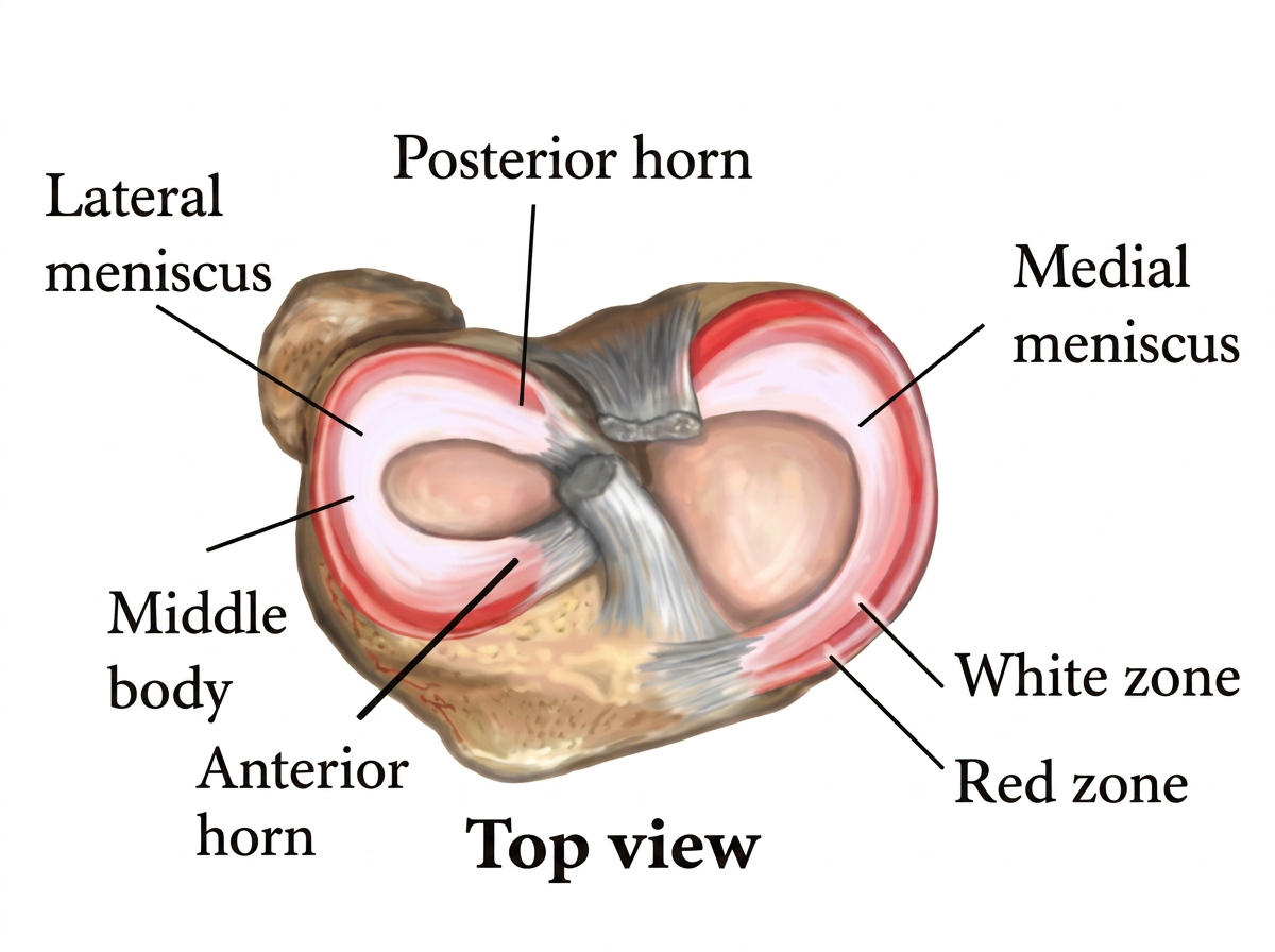
The Three Vascular Zones
The meniscus is divided into three zones based on blood supply:
Red-Red Zone (Outer Third)
The peripheral zone with the richest blood supply, fed by the medial and lateral geniculate arteries. Tears here have the best healing potential, spontaneously or following surgical repair. This is the zone where repair is most likely to succeed.
Red-White Zone (Middle Third)
A transitional zone where vascularity diminishes from the outer edge inward. Healing potential is variable. Surgical repair may be considered for appropriate tears in this zone, with outcomes less predictable than the red-red zone.
White-White Zone (Inner Third)
The avascular central portion. No meaningful blood supply exists here. Tears in this zone cannot heal spontaneously, and surgical repair is unlikely to succeed. Management is either conservative (for stable, asymptomatic tears) or partial meniscectomy (for symptomatic, unstable tears).
The vascular zone is the single most important factor in every meniscal treatment decision.
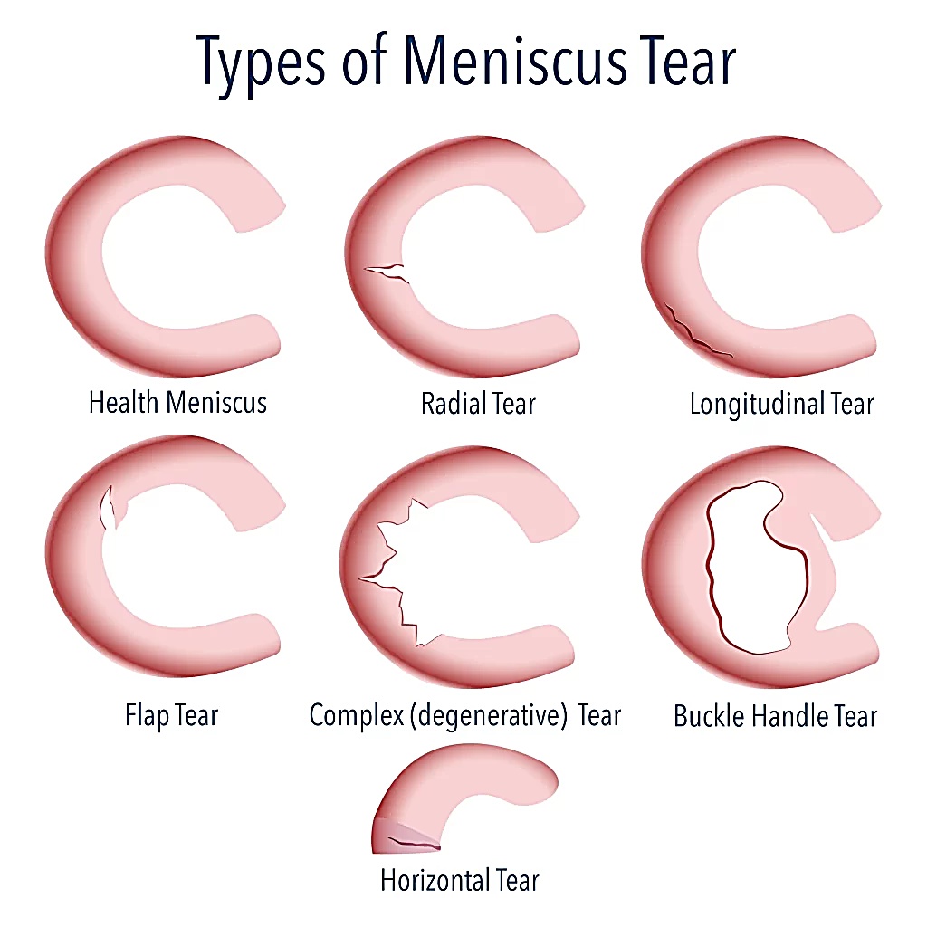
The Seven Tear Patterns
1. Vertical Longitudinal Tear
Runs parallel to the long axis of the meniscus. The most common traumatic tear pattern, particularly in younger athletes. Longitudinal tears in the red-red zone are the most amenable to surgical repair and heal better than other patterns in both operative and conservative management.
2. Bucket-Handle Tear
A full-thickness longitudinal tear where the inner fragment displaces into the intercondylar notch. Associated with a traumatic mechanism, often causes a "locked knee" with blocked extension. Typically requires surgical intervention. More common in younger patients and often associated with ACL injury.
3. Radial Tear
Runs perpendicular to the long axis, cutting across the circumferential collagen fibres. Mechanically disruptive, a complete radial tear abolishes the hoop stress function of the meniscus. Heals less reliably than longitudinal tears and complex radial tears in the white-white zone often require meniscectomy.
4. Horizontal Tear
Splits the meniscus into superior and inferior leaves. More common in middle-aged and older patients. Frequently associated with degenerative change and may present with a parameniscal cyst. Conservative management is typically first-line for horizontal tears.
5. Oblique (Flap) Tear
Combines vertical and horizontal elements, creating a mobile flap of tissue that can displace and catch within the joint. Can cause significant mechanical symptoms (clicking, catching, giving way) and may require surgical trimming if conservative management fails.
6. Complex Tear
Involves multiple planes and patterns, usually in the context of degenerative change. Most common in older patients. Conservative physiotherapy is the recommended first-line approach.
7. Root Tear
A tear at the anterior or posterior meniscal root attachments at the tibial plateau. Posterior root tears, particularly of the medial meniscus, are clinically significant because they abolish the hoop stress mechanism of the entire meniscus, accelerating cartilage loss. Often warrants surgical repair rather than conservative management.
Which Type Am I Most Likely to Have?
Younger athletes (under 35):
More commonly traumatic, vertical longitudinal, bucket-handle, or radial tears following twisting, pivoting, or contact injuries.
Active adults (35–55):
Mixed picture, may be traumatic or early degenerative. Horizontal and complex tears become more common.
Older adults (55+):
Predominantly degenerative lesions, horizontal, complex, or oblique tears in the context of broader cartilage change.
At IP Physio, we assess the full clinical picture alongside your imaging to determine exactly what type of tear you have and what it means for your management. Get in touch.
References
- Arnoczky SP, Warren RF. Microvasculature of the human meniscus. Am J Sports Med. 1982;10(2):90–95.
- Kopf S, et al. Meniscus tears: the good, the bad and the ugly, patterns classification and practical guide. World J Orthop. 2023. PMC10122773.
- Hammad M, et al. Meniscal tear repair: what's new in the literature? Transl Sports Med. 2025. doi:10.1155/tsm2/5511916.
- Maffulli N, et al. Treatment of meniscal tears: an evidence-based approach. World J Orthop. 2014;5(3):233–241. PMC4095015.
Still have questions?
Read quick, direct answers to the questions patients most commonly ask about meniscal tears.
Meniscal Tear: Frequently Asked Questions