


If a scan report has described your joint as "bone on bone", "degenerative", or "worn out", it's easy to hear that as a verdict on your future. It isn't. The relationship between what shows up on imaging and what you actually feel is far weaker than most people are told.
The Relationship Is Often Surprisingly Weak
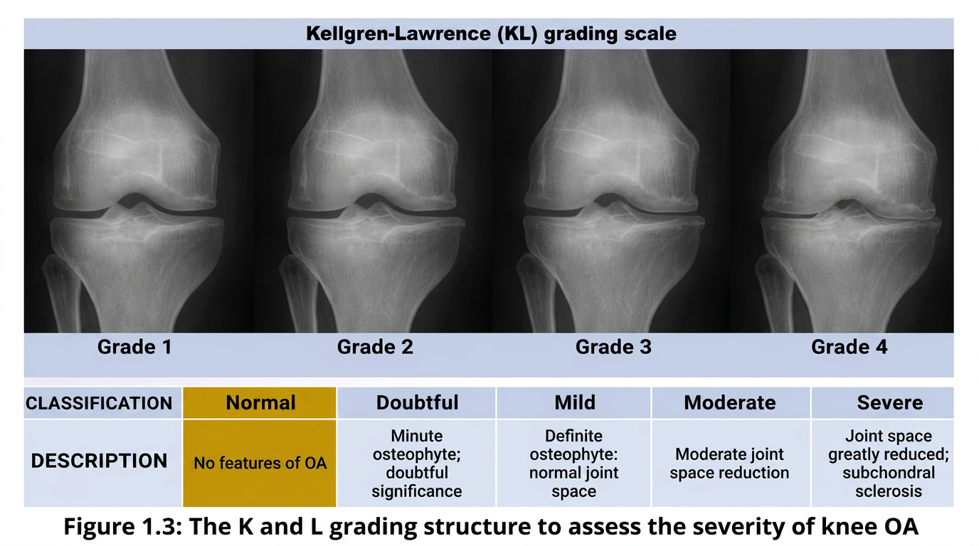
The relationship between the severity of osteoarthritis (OA) seen on X-ray and a person's symptoms is often surprisingly weak. Some people with severe structural changes on imaging have little or no pain. Others with relatively mild imaging findings have significant pain and disability. Structural findings, including cartilage thinning and osteophytes, are common in middle-aged and older adults with little or no joint pain.
Why Cartilage Loss Alone Doesn't Explain Pain
OA pain can arise from other innervated joint and surrounding tissues, including the subchondral bone, synovium, joint capsule and other periarticular tissues, rather than from cartilage itself. This is one reason the amount of cartilage loss visible on a scan doesn't map neatly onto how much pain someone experiences.
Changes in pain processing, including features of peripheral and central sensitisation, may also contribute to persistent or disproportionate pain in some people with OA. This is one reason that pain severity cannot always be predicted from the amount of structural change seen on imaging.
So What Should You Take From a Scan?
Imaging findings do not reliably predict the severity of symptoms in an individual person, and structural changes should always be interpreted alongside symptoms and physical function, not read in isolation. Imaging findings alone should not be used as a reason to stop moving or exercising.
NICE guidance reflects this: in people aged 45 or over with activity-related joint pain and no morning joint-related stiffness, or stiffness lasting no longer than 30 minutes, OA can usually be diagnosed clinically without imaging at all. Imaging is generally reserved for atypical presentations or when another diagnosis needs to be ruled out.
What This Means Practically
A "bone on bone" description is not a sentence, and it is not, on its own, a reason to avoid strengthening the muscles around the joint or to stop the activities you enjoy. Function, strength and how you're actually managing day to day are far more useful guides to what to do next than the wording on a radiology report alone.
Read More
Still have questions?
Read quick, direct answers to the questions people most commonly ask about osteoarthritis.
Osteoarthritis: Frequently Asked QuestionsReferences
- National Institute for Health and Care Excellence (NICE). Osteoarthritis in over 16s: diagnosis and management (NG226). 2022.
- Vincent TL. Peripheral pain mechanisms in osteoarthritis. Pain. 2020;161(9):S138–S146. doi:10.1097/j.pain.0000000000001923.
Get in touch with us today if a scan finding has left you unsure what to do next. A functional assessment can offer a clearer, more useful picture than the report alone.